Structure and Function of the Normal Heart
Before you begin to learn about heart disease, you must learn how the normal heart is constructed and how it functions. This is easier than you might think, because the heart is a surprisingly simple organ. An hour's easy reading will give you all the information you need to begin.
The Chambers of the Heart and their Connections
The heart is a hollow organ divided into four chambers, two on the top and two on the bottom (Fig. 1-1). Study this simple diagram until you know it as well as your own name: it's basic to everything else in the book.
The top two chambers are thin-walled structures that act primarily as holding chambers for the blood. They are called atria. This is the plural of the Latin word atrium, meaning “anteroom†or “porch,†and, in fact, these chambers do act as entryways to the great chambers below. The ventricles are large, thick-walled chambers that do the real work of pumping the blood. (This name comes from the Latin ventriculum, meaning a “cavity†or “pouch.â€)
Look again at Figure 1-1 and note the wall, or septum, that divides the left atrium from the right atrium and the left ventricle from the right ventricle. This wall of tissue is much like the septum in your nose that separates the two nostrils. The important thing to remember about the heart's septum is that it is absolutely watertight, or, more properly, “bloodtight.†Normally, no blood can pass through this septum from one side to the other. (It took the human race about 4,000 years to discover this simple fact. The ancient Greeks and Romans were convinced that blood somehow oozed through the septum from one side to the other. It doesn't.)
Physicians commonly refer to the right atrium and right ventricle together as the right heart and to the left atrium and left ventricle as the left heart.

The Motion of the Blood Through the Heart
The function of the heart can be described as a simple pump that forces blood forward by squeezing, in exactly the way that a bulb syringe forces out fluid when it's compressed.
The alert reader will at once ask, “If the blood doesn't flow from one side of the heart to the other through the septum, how does it ever move forward?†The answer to that question eluded philosophers and scientists until the English medical doctor William Harvey, in the early seventeenth century, discovered the simple circuit that is the basis of all modern cardiology.
The blood moves from the right heart to the left heart by way of the lungs. In other words, the right heart pulls the blood out of the veins and pumps it into the lungs. The left heart pulls the blood out of the lungs and pumps it on to the body.
(The outraged squalling of Harvey's contemporaries and the hoots of disbelief that greeted this profound truth are amusing to contemplate; they are also a little frightening.) Thus the heart and lungs together form a machine that takes oxygen out of the air, dissolves it in the blood, and pumps it to the tissues of the body.
Back to Structure: How Are the Heart and Lungs Connected?
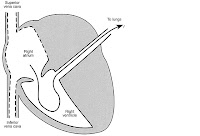
The blood that has completed its course through the tissues of the body flows back to the heart through the veins. The veins come together, growing larger, like streams combining into a river, until they end in two great veins that empty into the top and bottom of the right atrium. The word cava in Latin refers to something large or cavelike; hence the vein that empties into the top of the right atrium is called the superior vena cava, or, literally, “large top vein.†The great vein that empties into the bottom of the right atrium is logically called the inferior vena cava (Fig. 1-2). Blood flows from the right atrium down into the right ventricle and out to the lungs through the pulmonary artery (Fig. 1-3).

Within the lungs, the pulmonary artery branches into ever smaller arteries until it ends in a mass of capillaries—tiny vessels just wide enough to let one blood cell through at a time (Fig. 1-4). After the blood has been oxygenated it flows back to the heart through the four veins that empty into the left atrium (Fig. 1-5). Since these veins flow from the lungs to the heart they are called the pulmonary veins.


From the left atrium, blood flows down into the left ventricle and then out the aorta to the body (Fig. 1-6).
You must be thoroughly familiar with this circuit and with the names and function of the great vessels of the heart and lungs. The great vessels of the heart is the term used to include both arteries and veins.
Note: The great vessels of the heart are as follows:
The superior and inferior vena cavae, that empty all the blood from the body into the right atrium.
The pulmonary artery, which carries blood from the right ventricle to the lungs.
The pulmonary veins, which carry oxygenated blood from the lungs to the left atrium.
The aorta, or great artery, which carries the oxygenated blood out of the left ventricle to the body.
The Chambers of the Heart and their Connections
The heart is a hollow organ divided into four chambers, two on the top and two on the bottom (Fig. 1-1). Study this simple diagram until you know it as well as your own name: it's basic to everything else in the book.
The top two chambers are thin-walled structures that act primarily as holding chambers for the blood. They are called atria. This is the plural of the Latin word atrium, meaning “anteroom†or “porch,†and, in fact, these chambers do act as entryways to the great chambers below. The ventricles are large, thick-walled chambers that do the real work of pumping the blood. (This name comes from the Latin ventriculum, meaning a “cavity†or “pouch.â€)
Look again at Figure 1-1 and note the wall, or septum, that divides the left atrium from the right atrium and the left ventricle from the right ventricle. This wall of tissue is much like the septum in your nose that separates the two nostrils. The important thing to remember about the heart's septum is that it is absolutely watertight, or, more properly, “bloodtight.†Normally, no blood can pass through this septum from one side to the other. (It took the human race about 4,000 years to discover this simple fact. The ancient Greeks and Romans were convinced that blood somehow oozed through the septum from one side to the other. It doesn't.)
Physicians commonly refer to the right atrium and right ventricle together as the right heart and to the left atrium and left ventricle as the left heart.

The Motion of the Blood Through the Heart
The function of the heart can be described as a simple pump that forces blood forward by squeezing, in exactly the way that a bulb syringe forces out fluid when it's compressed.
The alert reader will at once ask, “If the blood doesn't flow from one side of the heart to the other through the septum, how does it ever move forward?†The answer to that question eluded philosophers and scientists until the English medical doctor William Harvey, in the early seventeenth century, discovered the simple circuit that is the basis of all modern cardiology.
The blood moves from the right heart to the left heart by way of the lungs. In other words, the right heart pulls the blood out of the veins and pumps it into the lungs. The left heart pulls the blood out of the lungs and pumps it on to the body.
(The outraged squalling of Harvey's contemporaries and the hoots of disbelief that greeted this profound truth are amusing to contemplate; they are also a little frightening.) Thus the heart and lungs together form a machine that takes oxygen out of the air, dissolves it in the blood, and pumps it to the tissues of the body.
Back to Structure: How Are the Heart and Lungs Connected?
The blood that has completed its course through the tissues of the body flows back to the heart through the veins. The veins come together, growing larger, like streams combining into a river, until they end in two great veins that empty into the top and bottom of the right atrium. The word cava in Latin refers to something large or cavelike; hence the vein that empties into the top of the right atrium is called the superior vena cava, or, literally, “large top vein.†The great vein that empties into the bottom of the right atrium is logically called the inferior vena cava (Fig. 1-2). Blood flows from the right atrium down into the right ventricle and out to the lungs through the pulmonary artery (Fig. 1-3).


Within the lungs, the pulmonary artery branches into ever smaller arteries until it ends in a mass of capillaries—tiny vessels just wide enough to let one blood cell through at a time (Fig. 1-4). After the blood has been oxygenated it flows back to the heart through the four veins that empty into the left atrium (Fig. 1-5). Since these veins flow from the lungs to the heart they are called the pulmonary veins.


From the left atrium, blood flows down into the left ventricle and then out the aorta to the body (Fig. 1-6).
You must be thoroughly familiar with this circuit and with the names and function of the great vessels of the heart and lungs. The great vessels of the heart is the term used to include both arteries and veins.
Note: The great vessels of the heart are as follows:
The superior and inferior vena cavae, that empty all the blood from the body into the right atrium.
The pulmonary artery, which carries blood from the right ventricle to the lungs.
The pulmonary veins, which carry oxygenated blood from the lungs to the left atrium.
The aorta, or great artery, which carries the oxygenated blood out of the left ventricle to the body.


Komentar